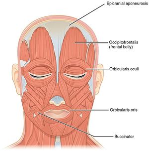
Located underneath the skin of the face and scalp are a group of 20 flat skeletal muscles. These muscles of facial expression, also named craniofacial muscles, are found in the subcutaneous tissue and emanate from bone or fascia, to attach onto the skin. They are a group of muscles that attach to skin and by contracting, the muscles pull on the skin and create movements of the face, such as smiling, grinning and frowning. Therefore, these muscles are commonly called muscles of facial expression, or mimetic muscles. All of the facial muscles are innervated by the facial nerve (CN VII) and vascularised by the facial artery.
The facial muscles are located around facial openings (mouth, eye, nose and ear) or extend over the skull and neck. Hence, they are divided into several groups;
- Muscles of the nose (nasal group)
- Muscles of the cranium and neck (epicranial group)
- Muscles of the external ear (auricular group)
- Muscles of the mouth or oral group (buccolabial group)
More specifically the oral group are accountable for movements of the mouth and lips. Such movements are necessary in singing and whistling and give emphasis to vocal communication.
Description
There is a total of eleven facial muscles that create movement at the mouth and their functions include:
- Lifting up and everting the upper lip: levator labii superioris, levator labii superioris alaeque nasi, risorius, levator anguli oris, zygomaticus major and zygomaticus minor muscles.
- Lowering and everting the lower lip: depressor labii inferioris, depressor anguli oris and mentalis muscles.
- Closing the lips: orbicularis oris muscle.
- Compacting the cheek: buccinator muscle.
The bulk of the mouth muscles are joined by a fibromuscular hub where their fibers insert. This structure is called the modiolus, it is founded at the angles of the mouth and it is primarily formed by the buccinator, orbicularis oris, risorius, levator anguli oris, depressor anguli oris and zygomaticus major muscles. The precise position of the modiolus varies, contributing to the variety of smile shapes which are seen in humans.
Each muscle is described individually below;
Orbicularis Oris
Origin
The fibres of the orbicularis oris surround the opening to the oral cavity. It comprises two parts; peripheral and marginal, with the border between them relating to the margin linking the lips and the surrounding skin. Both parts originate from the modiolus.
Insertion
- The peripheral part travels medially into the labial areas to insert on the dermis of the lips. In the midplane, a portion of the fibers intermingle with their respective fellow muscles to form the philtrum of the mouth.
- The marginal part travels from the modiolus on one side to the modiolus on the opposite side of the mouth. Some of the fibers curl upon themselves, forming the vermilion border, which is the boundary between the lips and the adjacent skin.
Nerve
Innervated by the buccal and mandibular branches of the facial nerve (CN VII).
Artery
Supplied from the superior and inferior labial branches of facial artery, with input from the mental and infraorbital branches of maxillary artery and the transverse facial branch of superficial temporal artery.
Function
A bilateral contraction of the whole muscle closes the mouth. An isolated contraction of particular parts of the muscle can produce various movements of the mouth, such as lip pouting, puckering, twisting and others. Through it's contractions the orbicularis oris assists speech and helps create a variety of facial expressions.
Assessment
Wearing a glove, place palpating fingers on the tissue of the lips. Ask the patient to pucker up the lips, and feel the contraction of the muscle. Once felt, palpate the entire muscle as the patient alternately contracts and relaxes.
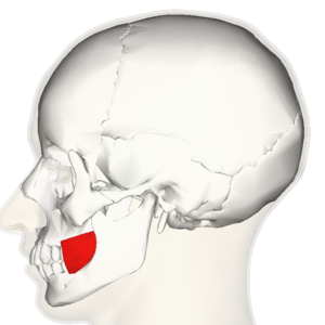
Buccinator
Origin
The buccinator muscle shapes the muscular structure of the cheek, filling the space between the maxilla and mandible. It is comprised of three parts; superior, inferior and posterior. It originates from the external lateral surface of the Alveolar process of maxilla, buccinator ridge of mandible, pterygomandibular raphe.
Insertion
All three parts of the buccinator coincide towards the angle of the mouth and insert onto the modiolus and mixes with other muscles of the upper lip.
Nerve
Buccal branch of facial nerve (CN VII)
Artery
Main supply from the buccal branch of the maxillary artery, with input from branches of the facial artery.
Function
The buccinator muscle compresses the cheek against the molar teeth and forestalls the cheek from getting bitten during mastication. Additionally, it aids to keeping the bolus of food centered in the oral cavity and limiting it from leaking into the oral vestibule. The buccinator has a role in playing wind instruments or whistling.
Assessment
Place palpating finger lateral and marginally superior to the corner of the mouth. Ask the patient to take a deep breath, purse lips, and press the lips on their teeth as if forcing air out while playing a trumpet to feel the contraction of buccinator. The majority of this muscle is deep to the masseter and other muscles of facial expression, therefore, palpation and it's specific identification is difficult.
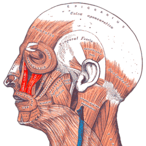
Levator labii superioris
Origin
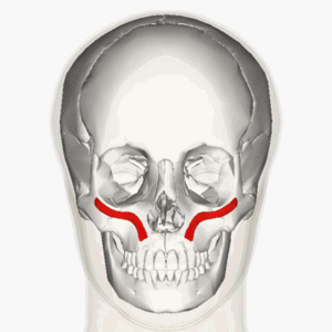
A short triangular muscle that originates from zygomatic process of maxilla and maxillary process of zygomatic bone.
Insertion
It travels inferiorly and medially to attach onto the skin and intermingle with the other muscles of upper lip.
Nerve
Zygomatic and buccal branches of facial nerve (CN VII).
Artery
Blood is supplied by the facial artery and infraorbital branch of the maxillary artery.
Function
This muscle aids other buccolabial muscles to lift and invert the upper lip, showing the maxillary teeth and deepening the nasolabial lines. This movement is necessary in making some facial expressions, such as snarling, smiling, grinning and contempt.
Assessment
Place palpating finger relatively half an inch or 1cm lateral to the center of the upper lip, at its superior margin. Ask the patient to lift the upper lip to show their upper gum and feel for the contraction. It can be challenging to identify the levator labii superioris from the close by levator labii superioris alaeque nasi (medially) and zygomaticus minor (laterally), as these also lift the upper lip.
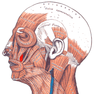
Levator labii superioris alaeque nasi
Origin
A slim, strap-like muscle found on both sides of the nose. It originates from the upper portion of the frontal process of the maxilla and travels inferolaterally.
Insertion
It inserts to the fascia and muscle tissue of the upper lip and the fascia and cartilage of the nose.
Nerve
Zygomatic and buccal branches of facial nerve (CN VII).
Artery
Facial artery and the infraorbital branch of the maxillary artery.
Functon
It elevates the lip and flares the nostril. This muscle is considered a muscle of facial expression of the nose and the mouth.
Assessment
Place palpating finger just lateral to the nose. Ask the patient to either lift their upper lip to show the upper gum or flare their nostril to feel the contraction. Again this muscle can be difficult to distinguish from close by nasalis (medially) and levator labii superioris (laterally).
Risorius
Origin
Risorius is inconsistent for it's point of origin. It may arise from assorted points that may subsume the fascia of the parotid gland, fascia of the masseter and platysma muscles, and periodically from the zygomatic arch.
Insertion
The fibers of the risorius gather medially and travel horizontally towards the angles of the mouth, where they mix with other facial muscles to form the modiolus.
Nerve
Supplied by the buccal branch of facial nerve (CN VII).
Artery
Superior labial branch of the facial artery.
Function
It's action is to pull the angles of the mouth laterally, and is often active during smile function.
Assessment
Place palpating finger closely to the corner of the mouth. Ask the patient to move the corners of the mouth laterally to feel the muscle contract. Ensure that the palpation is not too far superior on the zygomaticus major[4].
Levator anguli oris
Origin
A lean, sheet-like muscle that originates from the canine fossa of maxilla.
Insertion
It travels vertically inferiorly towards the angle of the mouth to insert on the modiolus, to mix with the other facial muscles.
Nerve
Zygomatic and buccal branches of facial nerve (CN VII)
Artery
Superior labial branch of facial artery and infraorbital branch of maxillary artery.
Function
Elevates the corners of the mouth and aids to create a smile, along with the risorius, zygomaticus major and minor.
Assessment
Place palpating finger directly superior to the corner of the mouth. Ask the patient to lift the corner of their mouth as if to show their canine tooth (a Dracual like expression) and feel for the contraction. The most superior portion of this muscle is deep to the zygomaticus minor and levator labii superioris and can be hard to distinguish. Additionally, is necessary to identify levator anguli oris from zygomaticus major, as both lift the corners of the mouth.
Zygomaticus major
Origin
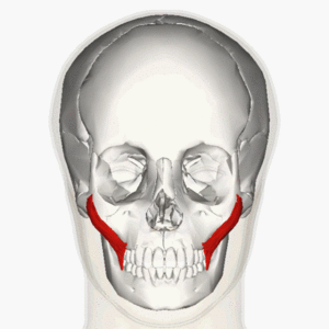
A slender muscle that originates from the lateral surface of the zygomatic bone.
Insertion
It travels diagonally to the angle of the mouth and donates to the disposition of the modiolus by mingling with several other facial muscles.
Nerve
Zygomatic and buccal branches of the facial nerve (CN VII).
Artery
Superior labial branch of the facial artery.
Function
It elevates the corner of the mouth and directs the corners laterally. It will aid to create a smile in cooperation with other muscles.
Assessment
Place palpating fingers directly superolaterally to the corner of the mouth. Ask the patient to smile to pull the corners of the mouth both superiorly and laterally to feel the contraction. It is challenging to determine zygomaticus major from close by levator anguli oris, and to distinguish it from zygomaticus minor that also lifts the upper lip.
Zygomaticus minor
Origin
Originates from the lateral surface of the zygomatic bone.
Insertion
Travels diagonally towards the lips. It attaches onto the skin of the upper lip, medial to the zygomaticus major.
Nerve
Buccal branch of facial nerve (CN VII),
Artery
Superior labial branch of facial artery.
Function
The zygomaticus minor works in unison with other tractors of the upper lip to lift and evert the upper lip. It facilitates several facial expressions including smiling and grimacing.
Assessment
Place palpating finger in estimation of half to three quarter inch (1-2cm) lateral to the center of the upper lip. Ask the patient to show you their upper gum to feel the contraction. This muscle needs to be differentiated from it's close neighbours, levator labii superiois (medially) and zygomaticus major (laterally).
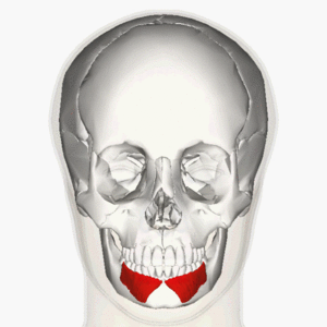
Depressor labii inferioris
Origin
A short quadrangular muscle located in the chin region. It attaches from the oblique line of mandible at the same time being continued with the labial part of the platysma.
Insertion
The muscle travels superomedially to attach on the skin and submucosa of the lower lip.
Nerve
Mandibular branch of facial nerve (CN VII).
Artery
Inferior labial branch of facial artery and mental branch of maxillary artery.
Function
It depresses, everts and draws the lower lip laterally. It also is responsible for pulling the lower lip inferomedially in conjunction with the labial part of platysma.
Assessment
Place palpating finger inferior to the lower lip and marginally laterally to the midline. Ask the patent to drop and draw their lower lip laterally to feel the contraction. It can be challenging to differentiate the depressor labii inferiois from close by depressor anguli oris, as both muscles drop and laterally move the lower lip/ corner of the mouth.
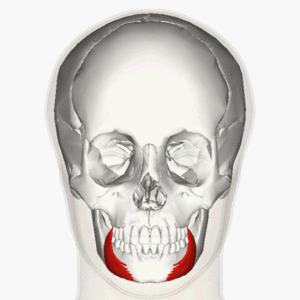
Depressor anguli oris
Origin
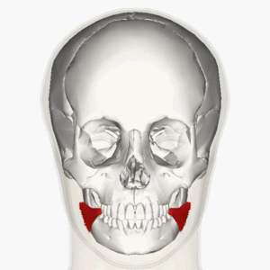
A triangular muscle located lateral to the chin on both sides of the face. It originates from the oblique line and mental tubercle of mandible.
Insertion
It travels in a nearly vertically upward direction to insert at the modiolus.
Nerve
Marginal mandibular and buccal branches of facial nerve (CN VII).
Artery
Inferior labial branch of the facial artery and the mental branch of the maxillary artery.
Function
Operates to drop the angle of the mouth, which assists to display feelings of sadness or anger. Furthermore, this muscle aids in opening the mouth during speaking or eating.
Assessment
Place palpating finger inferior and marginally lateral to the corner of the mouth. Ask the patient to frown by dropping and drawing the corner of the mouth laterally to feel the contraction of the muscle. Challenging to identify this muscle from close by depressor labii inferioris.
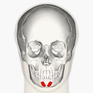
Mentalis
Origin
A short conical muscle found in the chin area. It originates from the incisive fossa of mandible.
Insertion
It travels inferiorly to insert on the skin of the chin at the level of the mentolabial sulcus of the mandible.
Nerve
Mandibular branch of the facial nerve (CN VII).
Artery
Inferior labial branch of facial artery and mental branch of the maxillary artery.
Function
Mentalis elevates the base of the lower lip, and in doing this it causes protrusion and eversion of the lower lip. These movements donate to certain actions such as shaping the lips when drinking, and creating facial expressions to display feelings of sadness, contempt and doubt.
Assessment
Place palpating finger relatively 1 inch (2.5cm) inferior to the lower lip and slightly lateral to the midline. Ask the patient to drop and protrude out the lower lip as if to pout and feel the contraction of the muscle. The inferior portion of mentalis is superficial and simple to palpate. The superior portion of the mentalis is more difficult to palpate and identify as it lies deep to depressor labii inferioris.
Clinical Relevance
Evaluation of the activity of the upper facial muscle group forms part of the assessment of Facial Palsy, which may be caused by Bell's Palsy, nerve damage in brain tumours such as Acoustic Neuroma (AKA Vestibular Schwannoma), Ramsay Hunt Syndrome, and other less common conditions.
Treatment
In similitude with all other muscles, the muscles of facial expression can become tight, particularly if the same facial expression is frequently made, demanding repeated contraction. When these small muscles become tight, they traction the covering fascia and skin toward its center, hence forming wrinkles that are directed perpendicularly to the path of the muscle fibers. Assessment of the pattern of facial wrinkles shows that wrinkles are perpendicular to the beneath muscle of facial expression. To stretch these muscles, it is requisite to move the face by creating a broad range of bold expressions. Each expression will stretch the muscles that produce the reverse expression. Therefore, the patient will have to make facial expressions they routinely do not make.


0 Comments