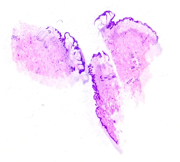
Skin biopsies are procedures done to investigate cutaneous growths or lesions. Many skin pathologies can be diagnosed by direct observation and palpation. However, there are others that require diagnostic procedures involving microscopic and histopathological examinations.
The various techniques that may be used to obtain skin biopsies include shave biopsy, punch biopsy and excisional biopsy. Skin biopsies, compared to other biopsies are relatively low-risk procedures and are associated with very few complications. However, like other biopsies, they require a consenting patient and anesthesia, typically local.

Shave Biopsies
Superficial shave biopsies are mainly used to evaluate lesions that are predominantly in the epidermis of the skin. These include skin tags, warts, and squamous or basal cell carcinomas. Under local anesthetic, a thin disk of tissue is removed for examination. The area is kept moist and covered for at least 7 days to prevent significant scar formation.This technique entails the use of a curved razor or small scalpel by an experienced surgeon to minimize the risk of scarring. It is the most commonly used technique. This is because it is relatively quick to perform, cost-effective and requires simple wound care. Shave biopsies may be done superficially, or via saucerization, and produce flat specimens.
Saucerization biopsies require the use of curved razor blades to obtain specimens from lesions that extend deeper into the skin, that is, into the dermis and subcutaneous fat. Indications for saucerization biopsies include suspicious lesions that are heavily pigmented or lesions that are difficult to remove via the superficial technique due to their anatomical location. Moreover, saucerization is preferred in the case of lesions in anatomical locations that are likely to produce scars that become hypertrophic or spread excessively. These include those found on the ears or upper arm.
Punch Biopsies
Punch biopsies produce cylindrical specimens due to their circular blades that are rotated through the skin to the subcutaneous tissue. These biopsies may be incisional or excisional. The type of punch biopsy adopted depends on the type of tissue and the size of the lesion. Lesions biopsied with this method include dysplastic nevi and bullous or inflammatory eruptions. These biopsies may require sutures to assist with quicker wound healing and to improve the chances of a better cosmetic result. Despite their ability to provide full-thickness specimens, punch biopsies are limited in terms of the width of sample obtained. This limitation is particularly crucial in tumor staging and in arriving at a prognosis of malignant lesions.
Excisional Biopsies
These biopsies involve removal of the entire lesion, including a margin of surrounding healthy skin as well. They are ideally suited for the removal and diagnosis of small melanomas. Excisional biopsies require sutures and may heal with scarring. Skin grafts or flaps may be used to replace large areas of skin that may have been removed during the procedure.


0 Comments